Isotype of this species is deposited in the Croatian National Diatom Collection as permanent slide under accession number HRNDC000007.
From the Latin word sulcata (‘ploughed’), with reference to the external cord-like silica strips and grooves on the valve face.
Marine Turtle Rescue Centre, Pula, Croatia (44°50′07′′N, 13°49′58′′E). Collected from a semi-adult (45 kg) female loggerhead turtle Caretta caretta named ‘Palma Modesty’ by K. Gobić Medica, 30 September 2016 (holotype).
Kosi Bay, South Africa (26°59′39′′S, 32°51′60′′E). Collected from the carapace of the adult female loggerhead C. caretta (tag numbers: ZA0762D, ZA0763D) by R. Majewska, 15 December 2017 (paratype).
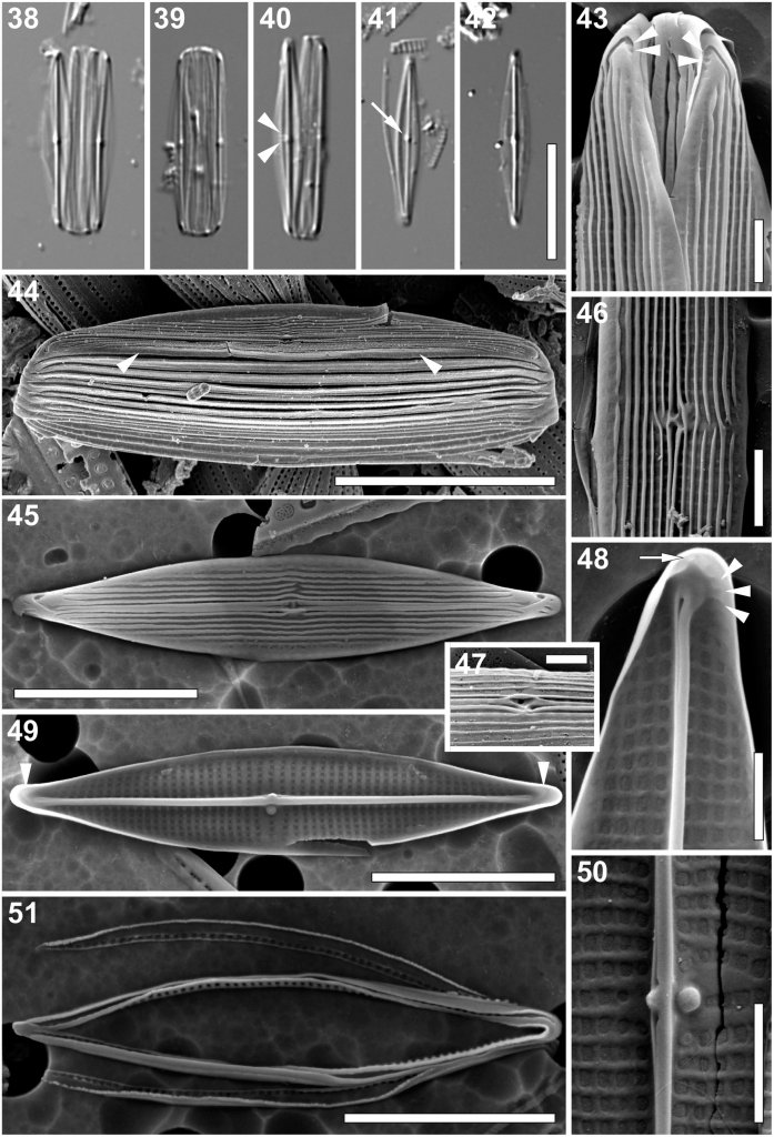
Light microscopy (Figs 38–42):
Frustules in girdle view broad with numerous girdles bands (Figs 38–40). Valves narrowly lanceolate with elongated acutely rounded to very slightly rostrate apices (Figs 38–42). Valve dimensions (n = 30): length 10–28 μm, width 2.5–4 μm, length/width ratio 4.0–7.4. Axial area narrow, with prominent raphe-sternum (Figs 38–42). Central area barely distinguishable, with two central striae more widely spaced and visible (Fig. 40, arrowheads). Other striae indiscernible. Fistula visible as glistening dot in the central area (Fig. 41, arrow). Girdle bands open, appearing unornamented (Figs 38–41).
Scanning electron microscopy (Figs 43–51):
External view: Valve face flat. Several distinct longitudinal cord-like silica strips running from apex to apex. Near the axial area and close to the valve face/mantle junction, strips occasionally interrupted (Figs 43–47). Mantle shallow, unperforated. Very narrow pore-free border running entirely around the valve at the valve face/mantle junction (Fig. 44, arrowheads). Striae uniseriate, barely visible between the longitudinal silica strips, parallel to weakly convergent at the apices (Figs 43, 46), 40–45 in 10 μm. Areolae rectangular, more or less uniform in size (Figs 43, 46, 47). Raphe branches straight, running between two raised silica ridges developing into a very narrow conopeum-like structure (Figs 43, 45–47). Central raphe endings straight, slightly expanded, with raised silica rims creating spatulate openings, clearly deflected towards the secondary side of the valve (away from the fistula; Figs 45–47). Fistula mostly hidden beneath the pocket-like thickening (formed by the fusion of longitudinal silica strips) with linear opening parallel to the raphe-sternum (Figs 45–47). Terminal raphe fissures strongly hooked towards the secondary side of the valve (Figs 43–45), with up to three small areolae at the end of the fissure (Fig. 43, arrowheads). Several apically elongated to irregularly shaped areolae present at the valve apex perpendicular to the valve margin (Fig. 45).
Internal view: Striae parallel becoming slightly convergent towards the apices (Figs 48–50), composed of square areolae, occluded by perforated hymenes. Areolae uniform in size across the valve face, only slightly larger and slightly raised close to the raphe sternum (Figs 48–50). Fistula occluded by one domed hymen forming a ball-like structure, not reaching the raphe-sternum (Figs 49, 50). Raphe slit opening laterally onto the uniformly thick raphe-sternum towards the secondary side (Figs 48–50). Central raphe endings very slightly expanded, slightly deflected towards the fistula (Figs 49, 50). A lateral and slightly raised rounded or nodular thickening present on the raphe-sternum, between the central raphe endings (Figs 49, 50). Terminal raphe endings straight, lying somewhat laterally on the sternum, terminating onto simple helictoglossae (Figs 48, 49). Thickenings corresponding to the hooked external terminal raphe fissures present poleward from the helictoglossae (Figs 48, 49, arrowheads). Areolae at the apices (Fig. 48, arrow) and those at the end of the curved thickenings (Fig. 48, arrowheads) occluded by slightly raised hymenes. Cingulum composed of multiple open copulae (Figs 44, 51). Pars interior with a row of hymenate, rectangular pores (Fig. 51); ca. 6 in 1 μm. Pars exterior plain (Fig. 44).